What Is 3D Imaging And How Is It Changing The World Of Dentistry?
3D imagingis a technology that provides the illusion of depth inside a picture by converting 2D data into a 3-dimensional format.
Stereography is used in 3D imaging, as we can see from a familiar source: the human visual system.
Humans perceive with two eyes that are somewhat apart.
This enables individuals to experience depth in addition to the horizontal and vertical information represented by a normal 2D television screen, for example.
Three-dimensional technologies are one of the most recent and essential advances in dentistry.
These systems, including intraoral scans, 3D imaging tests (CAT scan, CBCT, and MRI), CAD/CAM 3D printing devices, and 3D computer software, have allowed clinicians to improve patient care while shortening treatment planning time significantly.
The overall improvement provided by these technologies can improve clinicians' workflow and effectiveness by simplifying traditionally tricky techniques.
3D Imaging In Orthopedic
Orthodontic and dentofacial orthopedic diagnosis and treatment planning rely on imaging, jaw monitoring, and functional evaluations.
These approaches recreate or explain anatomical and physiological facts and depict 3D anatomy.
Orthodontists use imaging to analyze and record craniofacial size and shape. Orthodontists employ 2D static imaging to capture craniofacial anatomy. However, the depth of structures cannot be determined.
3D imaging was created in the 1990s and is used in orthodontics, dentistry, and orofacial surgery.
In 3D diagnostic imaging, anatomical data is obtained, processed by a computer, and shown on a 2D monitor to create the appearance of depth.
Orthodontics and orthognatic surgery focus on facial soft and hard tissues and teeth. The trio helps arrange orthodontic therapy.
Imaging these structures helps physicians decide on a therapy method.
3D imaging for orthodontics includes pre and post-treatment examination of dentoskeletal and craniofacial connections, facial appearance and beauty, and 3D treatment predictions.
Using 3D imaging in orthodontics also helps make diagnostic choices and arrange therapy.
3D Imaging History
Thalmann-Degan documented facial differences after orthodontic treatment in 1944. This was the first clinical study of stereophotogrammetry.
Parallel to computer developments, computerized stereophotogrammetry has entered the market and has provided faster, more comprehensive, and correct sequence taking and construction.
The 3D imaging technique has been enhanced for use in various areas of health sciences.
Stereophotogrammetry has been introduced to provide more extensive and accurate assessments of the captured things by improving old photogrammetric techniques.
A 3D model can be built and monitored from any perspective and measured from any direction using one or more converging pairs of views.
Around 40 years ago, the first CT scanning device was developed.
After a short period, a stack of CT sectional images was used to obtain 3D data.
Clinicians began using 3D imaging in craniofacial deformities in the early 1980s.
In 1986, the first simulation software for craniofacial surgical needs was introduced.
3D CT and MRI principles and applications in medicine were then published.
A distinct discipline in 3D imaging was established, dealing with various types of imaging, manipulation, and analysis of multidimensional medical structures.
3D Imaging Techniques
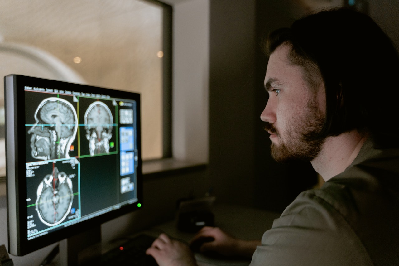
- Computed Tomography:Computed tomography (CT) imaging creates body cross-sections using X-rays. The CT scanner scans a patient's body with a narrow, fan-shaped X-ray beam. Specific tests require a contrast substance to make certain body parts more visible, but the procedure is painless. Advanced fan-beam CT can simultaneously achieve 64 and 128 sections.CT scans provide 3D data on soft and hard tissues. Comparatively, CT data is inadequate for soft-tissue imaging.
- Cone Beam Computerized Tomography (CBCT): Craniofacial CBCT devices aim to improve traditional CT scanning. Traditional CT scanners emit 15 times more radiation. CBCT realigns 2-D images in coronal, sagittal, oblique, and other planes.CBCT's radiation dose equals 12 panoramic radiographs. CBCT organizes 3D data on PCs. Implant and orthodontic software are advanced. CBCT can show soft tissues and hard head, and facial tissue. CBCT increased the incidence of oral abnormalities, according to previous research.
- The Nasopharyngeal Airway Analysis:CBCT has improved nasopharyngeal airway analysis. If necessary, adenoids/tonsils can be surgically removed, or obstructive sleep apnea therapy can be used. After orthodontic treatment, airways were unchanged in both studies.
- Cleft Lip/Palate Patients (CL/P Patients): CBCT is necessary for congenital abnormalities. Studies on CBCT imaging in orofacial monsters have focused on CL/P because it's so common. CBCT volumetric studies can determine the right amount of graft material for CL/P patients. Synchronous rhinoplasty is recommended based on CBCT soft tissue measurements across the three groups to improve nasal and labial appearance.
- Temporomandibular Joint (TMJ) Morphology:Images of cranial structures from different periods can be placed using 3-D software. CBCT images can show facial changes caused by tooth movement, orthognathic surgery, or craniofacial therapy. 3D Fotoscan machines can create CBCT models.
- Micro-Computed Tomography (MCT): MCT is like CT, but the reconstructed cross-sections are more minor. CT scans capture 0.012 mm thin cross-sections, but MCT can capture nano-sized sections. Non-invasive MCT analyzes mineralized tissues. MCT's future lies in its ability to collect input over a smaller volume than the whole body, minimizing radiation exposure.
- 3D Laser Scanning:Laser scanning provides 3D images for treatment planning or analyzing the results of orthodontic and orthognathic therapy. 3D laser scanners create digital models.
- Structured Light Technique: Structured light scanning allows non-ionizing 3-D face contouring. Ora-Scanner, the first handheld 3D intra-oral scanner, uses structured light. Techalertpaisan and Kuroda created a 3D face form using two LCD projectors, a camera, and a computer. This technique takes at least 2 seconds to take a picture, which may be too long for babies and toddlers.
- Sterophotogrametry:Stereophotogrammetry involves photographing a 3-D object from two planes. This strategy works well for face display. Thalmann-Degan used stereophotogrammetry to record orthodontic changes in 1944. Tissue reflections, hair and brow interference, changing posture between views, and motions during imaging reduce the accuracy of 3D face photos.
- 3D Facial Morphometry (3DFM):The system includes two infrared cameras, marker detection hardware, and 3D landmark reconstruction software. Landmark placement is labor-intensive and time-consuming. This technique can't create facial expressions with soft tissue.
- Magnetic Resonance Imaging (MRI): Magnetic fields direct radio waves to the analysis spot. MRI uses a hydrogen resonance signal. Radio stimulated cells to convert hydrogen energy into numbers. Computer processing creates images.
Conclusion
3D imaging tools in orthodontia are critical in providing more investigative evidence on specific situations, such as individuals with craniofacial incongruities.
Overall, if these 3D imaging modalities become standard practice for orthodontists, the chair time for complete dental and oral records, record loss, and storage of these dental and oral data will be decreased, potentially increasing the knowledge base for multidisciplinary communication.
Although evidence-based standards for 3D imaging are still required before it can be used in routine dentistry, in the long run, 3D imaging provides health practitioners with a dynamic 4D virtual patient in motion.
People Also Ask
What Is The Benefit Of 3D Medical Imaging Compared To 2D?
It has also been said that 3D has an advantage over 2D because computerized 3D pictures may be interactive, enabling the viewer to gain information from their motions or controls.
It can also employ elements like music, contributing to the viewer's 'immersion' in the image.
Why Is 3D Medical Imaging Important?
3D visuals improve knowledge and enable surgeons to plan more precisely before an operation.
Even under the best of conditions, medical skill is dispersed around the globe.
What Is The Difference Between 2D And 3D Images?
2D is "flat," employing the horizontal and vertical (X and Y) dimensions; the picture has only two dimensions and becomes a line when rotated to the side.
The depth (Z) dimension is added in 3D. This third dimension enables rotation and viewing from various angles.